de winters ecg pattern
The de Winter electrocardiogram pattern is a transient electrocardiographic phenomenon that presents at early stage of ST-segment elevation myocardial infarction Clin Cardiol 41 2018 pp. LAD occlusion can present de Winter ST-T changes.
De Winter St T Waves Ecg Medical Training
However it is often unrecognized by physicians.
. The De Winter ECG pattern was reported as an indicator of acute left anterior descending LAD coronary artery occlusion and is considered an anterior ST-elevation myocardial infarction STEMI equivalent1 The key diagnostic features include ST-depression and peaked T-waves in precordial leads and it can be seen in around 2 of patients with anterior myocardial. 1 The ECG reveals a de Winters pattern which shows a 1. Cardiac magnetic resonance imaging confirmed the diagnosis of my.
First reported by Dutch Professor of Cardiology Robbert J. An electrocardiographic finding suggestive of impending myocardial infarction the de Winters pattern or de Winters T-waves describes an abnormality thought to be indicative of acute occlusion of the proximal left anterior descending coronary artery LAD 2. 1 BACKGROUND Winter et al 2008 described an ECG pattern without ST-segment elevation in ECG leads which consisted of a 13 mm upsloping ST-segment depression at the J-point in precordial leads V1-V6 followed by tall positive symmetrical T-waves and ST elevation of 12 mm in lead aVRThe de Winter ECG pattern known as a STEMI equivalent has a high predictive.
Earlier percutaneous coronary intervention PCI reduces. Emergent coronary angiography was used to rule out the presence of significant stenosis. Onset to recording the de Winter pattern was 99 79 min.
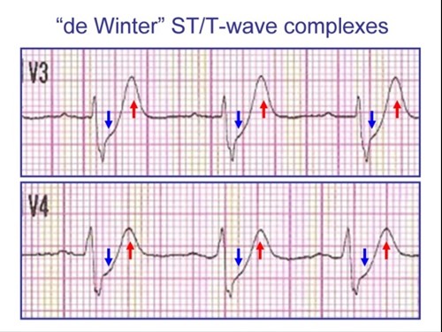
The main characteristics of the de Winter electrocardiogram ECG pattern are up-sloping ST-segment depression in the V 1 to V 6 leads followed by tall and symmetrical T waves which remain consistent with no evolutionary ECG changes. We present a patient with acute proximal LAD occlusion who presented with an evolutionary ECG change in which the de. The maximal amplitude of the STD was 3 2 mm.
The De Winter ECG pattern has been reported to indicate acute left anterior descending coronary artery occlusion and is often considered to be an ST elevation myocardial infarction STEMI equivalent. The de Winter ECG pattern is a recently-described STEMI equivalent that emergency physicians and paramedics must be aware of. De Winter in 2008 the de Winter ECG pattern is an anterior STEMI equivalent that presents without obvious ST segment elevation.
The reported positive predictive value PPV for the de Winter ECG pattern to predict an acute left anterior descending artery LAD lesion is inconsistent. We show that STsegment. The main characteristics of the de Winter electrocardio-gram ECG pattern are up-sloping ST-segment depres-sion in the V 1 to V 6 leads followed by tall and symmetrical T waves 1 which remain consistent with no evolutionary ECG changes.
These patients are suffering occlusion myocardial infarction OMI and require immediate reperfusion therapy. The de Winter ECG pattern occurred after ST-segment elevation in a patient with chest pain Intern Emerg Med. The de Winter electrocardiograph ECG pattern in patients with chest pain is associated with occlusion of the proximal left anterior descending LAD artery.
We aimed to investigate the morphology of the De Winter ECG pattern and evaluate the test characteristics of the De Winter pattern for the diagnosis of acute coronary occlusion. The de Winter sign is a rare electrocardiogram ECG manifestation of proximal LAD occlusion. De Winter syndrome is a rare electrocardiographic ECG pattern that makes the diagnosis of ST-segment elevation myocardial infarction STEMI very challenging.
Recently several case reports have indicated that this ECG pattern may evolve into ST-elevation myocardial infarction. The maximal amplitude of the positive tall T wave was 9 3 mm. Besides the morphology of upsloping or nonupsloping ST depression STD may have different significance of severity and prognostication.
1 The previous view was that the de Winter ECG pattern is static. 1 ST-segment elevation myocardial infarction STEMI equivalent patterns make the diagnosis of STEMI very challenging. 1 It is thought to be related to acute anterior descending artery occlusion.
The de Winter electrocardiographic ECG pattern was characterized by upsloping ST-segment depressions tall and positive symmetrical T waves in precordial leads. This review article focused on 5 under recognized high-risk ECG patterns in the ACS patient that result in poor outcomes including malignant dysrhythmias higher rates of. Timely recognition of this pattern is crucial.
It has become increasingly recognized as an STsegment elevation myocardial infarction equivalent pattern due to proximal left anterior descending pLAD artery occlusion. These patients typically have critical stenosis of the LAD requiring emergent PCI or thrombolysis. De Winter sign in ECG is one of the not so uncommon types of presentation for acute anterior wall myocardial infarction.
These patterns were once considered stable conditions without dynamic evolution of ECG. The heart rate when recording the ECG with de Winter pattern was 74 18 bpm. We present a patient with.
Our case indicates that early identification and diagnosis of such ECGs and timely reperfusion therapy of De Winter syndrome as an ST-segment elevation myocardial infarction STEMI equivalent are. The exact mechanism for this ECG pattern is not known. The De Winter ECG pattern has been reported to indicate acute left anterior descending coronary artery occlusion and is often considered to be an ST elevation myocardial infarction STEMI equivalent.
Table 1 shows the ECG and angiographic findings of the two groups. However it is often missed by physicians as well as cardiologists leading to less aggressive initial management. There are also STEMI equivalent patterns that are caused by occlusion of the coronary arteries that place a significant portion of the left ventricle at jeopardy and result in poor outcomes.
This rare ECG pattern was recognized as an indication of proximal left anterior descending artery occlusion. We aimed to investigate the morphology of the De Winter ECG pattern and evaluate the test characteristics of the De Winter pattern for the diagnosis of acute coronary occlusion. De winter syndrome is a rare pattern that can occur on an electrocardiogram ECG.
In 2008 de Winter et al described an ECG pattern suggesting that it should be considered an ST-elevation myocardial infarction STEMI equivalent de Winter Verouden Wellens Wilde 2008 with the potential to predict critical stenosis or occlusion of the left anterior descending coronary artery LAD. De Winter electrocardiograph ECG pattern signifies proximal left anterior descending coronary artery LAD occlusion and extensive anterior myocardial infarction and it is found in about 2 of patients with proximal LAD occlusion. A 26-year-old man presented to the emergency department with chest pain and electrocardiogram ECG changes compatible with the de Winter pattern.